

 Medically Reviewed
ECHO vs ECG: Which Heart Test Do You Actually Need?
When a doctor orders a heart test, two names come up most often: ECHO and ECG. Both are non-invasive and widely used in cardiac care. But they measure entirely different things and answer very different clinical questions. Understanding the difference between ECG and echo helps you walk into your appointment informed — and helps you understand why one test may not be enough on its own.
Key Takeaways
- An ECG checks the heart’s electrical signals. It can help find problems when someone has chest pain, a fast heartbeat, or an irregular heartbeat.
- An ECHO takes pictures of the heart. It shows how the heart looks and how well it is working.
- Research shows that ECG has limited sensitivity in detecting structural changes such as left ventricular hypertrophy, making ECHO the more reliable tool in such cases.
Quick Answer: An ECG checks heart rhythm, whereas an ECHO checks heart structure. A doctor may order one or both, depending on your symptoms.
- What Is an ECG?
- What Does an ECG Detect?
- What an ECG Cannot Do
- What Is an Echo?
- What Does an Echo Detect?
- ECHO vs ECG: The Main Difference
- When Does a Doctor Order an ECG?
- When Does a Doctor Order an Echo?
- When Both Tests Are Needed Together
- Why ECG Alone Is Not Enough for Detecting Structural Heart Changes
- What to Expect During Each Test
- ECG or Echo: Which Is Better?
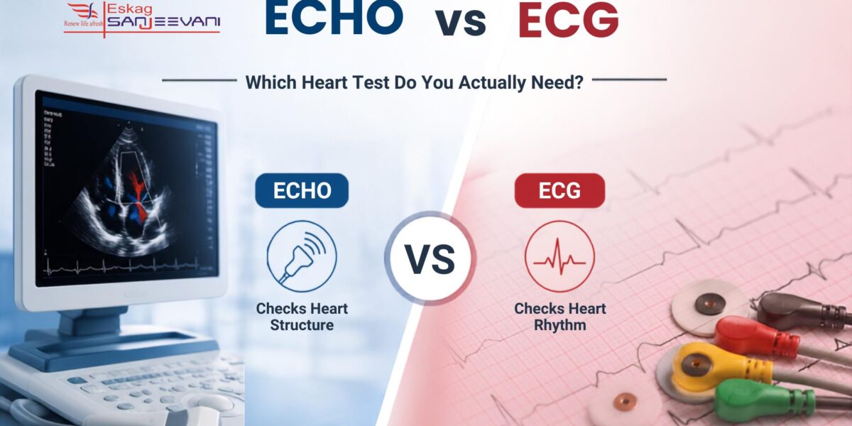
What Is an ECG?
An ECG or electrocardiogram is a simple and painless test that checks your heartbeat. Small adhesive electrode patches are placed on your chest, arms, and legs and connected to an ECG machine by lead wires. The machine then measures, interprets, and prints out your heart’s electrical signals. No electricity is sent into the body.
With every heartbeat, an electrical impulse travels from the top of the heart downward, prompting the chambers to contract and pump blood. The ECG captures this impulse as a waveform on paper or screen. A standard 12-lead ECG uses 10 physical electrodes to generate 12 different views of the heart, 6 precordial (V1–V6) and 4 limb electrodes, giving cardiologists a comprehensive picture of how electrical signals propagate through the heart muscle.
The whole procedure takes under 10 minutes. At Eskag Sanjeevani, our ECG test in Kolkata is part of our dedicated cardiac diagnostics programme, with every result reviewed by an experienced cardiologist.
What Does an ECG Detect?
An ECG is the primary test for electrical and rhythm-based heart problems. It can detect conditions before they cause structural changes visible on imaging. Some of the main ECHO vs ECG distinctions start here: the ECG’s value lies in rhythm, not structure.
Conditions an ECG can identify include:
- Atrial fibrillation and other arrhythmias that cause the heartbeat to become irregular, too fast, or too slow.
- Signs of a heart attack, including characteristic ST-segment elevation or depression on the tracing.
- Conduction blocks are where electrical signals are delayed or interrupted between the upper and lower chambers.
- Electrolyte disturbances such as abnormal potassium or magnesium levels that disrupt heart rhythm.
- Baseline indicators of hypertension-related heart changes, though with limited diagnostic reliability [1].
What an ECG Cannot Do
An ECG only captures electrical data. It cannot show the physical structure of your heart, its size, wall thickness, valve function, or pumping efficiency. If your doctor suspects a structural problem, an ECG will not provide a complete answer. This is the key limitation that makes the ECG vs echo comparison so clinically important.
What Is an Echo?
An ECHO or echocardiogram is an ultrasound scan of the heart. It uses high-frequency sound waves to produce real-time moving images of your heart, showing how it looks and functions at the structural and mechanical levels.
The most common type is the transthoracic echocardiogram (TTE), in which a small ultrasound probe is placed on the chest. Other types include:
| Type of ECHO | How It Is Done | Primary Use |
| Transthoracic (TTE) | Probe on chest surface | General heart structure and function |
| Transesophageal (TEE) | Probe passed into oesophagus | Detailed valve assessment, clot detection |
| Stress ECHO | TTE performed during or after exercise | Detecting artery blockage under exertion |
| Fetal ECHO | Probe on mother’s abdomen | Assessing heart structure in unborn babies |
At Eskag Sanjeevani, our cardiology department offers echocardiography alongside the full range of cardiac investigations, including 24-hour Holter monitoring and CT coronary angiography.
What Does an Echo Detect?
An ECHO provides detailed information about the heart’s anatomy and mechanical performance. It is the reference standard for diagnosing structural heart conditions. Key things an ECHO can assess include:
- Left ventricular ejection fraction — how much blood the heart pumps out with each contraction- is a key measure of heart failure.
- Valve structure and function, including stenosis (narrowing) and regurgitation (leaking) of any of the four valves.
- Left ventricular wall thickness and mass are used to diagnose or rule out left ventricular hypertrophy (LVH).
- Pericardial effusion is fluid accumulation around the heart that can compress it and affect function.
- Congenital structural defects such as septal holes or abnormal vessel connections are present from birth.
ECHO vs ECG: The Main Difference
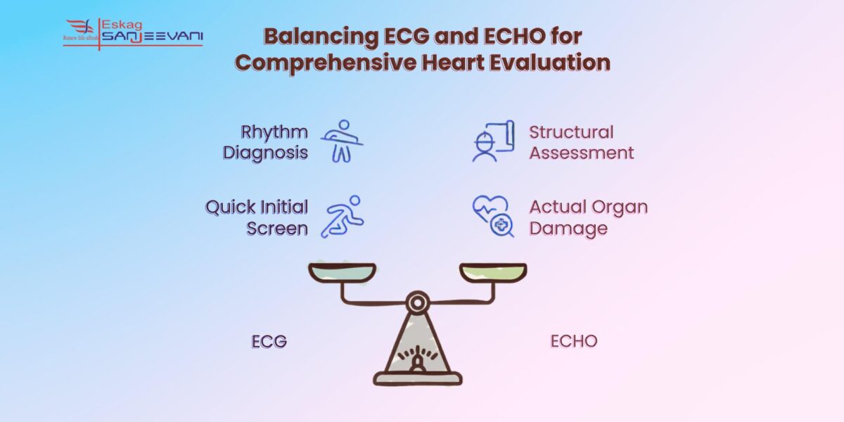
An ECG and an ECHO do different things. An ECG checks the heart’s electrical signals. Whereas an ECHO shows the heart and how it moves. So, one test cannot do the job of the other.
| Feature | ECG | ECHO |
| What it measures | Electrical activity | Structure and pumping function |
| Technology used | Electrodes and lead wires | Ultrasound waves |
| Duration | 5–10 minutes | 20–45 minutes |
| Best for | Arrhythmias, heart attacks, and conduction disorders | Valve disease, heart failure, LVH, cardiomyopathy |
| Can it show the heart structure? | No | Yes |
| Can it detect rhythm problems? | Yes | Limited |
| Radiation involved? | No | No |
| Is it painful? | No | No |
When asking which is better, ECG or echo, the answer depends on what your cardiologist needs to find. Neither test is universally superior; they serve different diagnostic purposes.
When Does a Doctor Order an ECG?
An ECG is typically the first cardiac test ordered in an emergency or outpatient setting because it is fast, inexpensive, and widely available. Your doctor is likely to recommend an ECG for:
- Chest pain that moves to your arm, jaw, or back. An ECG may be done to check if the pain is linked to a heart attack.
- Palpitations, racing heartbeat, or irregular beats that suggest an arrhythmia.
- Unexplained dizziness or fainting episodes, which can result from serious electrical disturbances.
- Feeling out of breath or tired after doing something active.
- Pre-surgical screening and routine cardiac risk assessment for high-risk individuals.
Also read: Chest Pain That Comes and Goes: 8 Causes That Aren’t a Heart Attack
When Does a Doctor Order an Echo?
An ECHO is ordered when the clinical concern relates to the heart’s structure or pumping ability — not just its rhythm. Your doctor is likely to recommend an ECHO for:
- Suspected or confirmed heart failure, to measure ejection fraction and understand its cause.
- Known or suspected valve disease, to assess severity and guide treatment decisions.
- Monitoring hypertension-related heart damage, where left ventricular hypertrophy is a concern.
- Cardiomyopathy — abnormal thickening, enlargement, or stiffening of the heart muscle.
- Further investigation after an abnormal ECG result is needed to check whether structural damage has occurred.
For patients already on a TMT stress test pathway, an ECHO often follows to confirm structural findings.
When Both Tests Are Needed Together
Some conditions affect both electrical function and heart structure. In those cases, your cardiologist may order both an ECG and an ECHO as part of the same evaluation.
A patient presenting with atrial fibrillation, for example, would have an ECG to confirm the rhythm diagnosis and an ECHO to check whether the fibrillation has affected chamber size or pumping function. Similarly, a patient with high blood pressure may have both tests, the ECG as a quick initial screen and the ECHO to assess actual organ damage.
Why ECG Alone Is Not Enough for Detecting Structural Heart Changes
This is one of the most important findings in recent cardiac research, and a key reason the ECG vs echo conversation matters clinically.
A 2024 study looked at 329 people who had high blood pressure. The researchers used both an ECG and an ECHO to check if the left side of the heart had become enlarged. This condition is called left ventricular hypertrophy (LVH). The ECHO confirmed LVH in 70 people, which was 21.3% of all the people in the study. The researchers then checked how well an ECG could find the same problem. The ECG found 47.9% of the people who had LVH. Its specificity was 75.3%. The area under the ROC curve was 0.604, which also showed that the ECG did not work very well for finding LVH. The study found that a standard ECHO is a more reliable way to check for this heart problem [2].
A separate longitudinal analysis from the PAMELA study followed 927 participants over approximately 10 years, examining whether ECG criteria could track changes in left ventricular mass (LVM) over time, with echocardiography as the reference standard. The findings showed that as ECHO-confirmed LVH prevalence rose from roughly 10% at baseline to around 30% at follow-up, the most commonly used ECG criteria — Sokolow-Lyon and Cornell voltage showed a further reduction in sensitivity over time. ECG variations were also found to move in the opposite direction of ECHO-measured LVM changes in most subjects, leading the authors to conclude that ECG is unsuitable for the longitudinal evaluation of LVM changes [1].
Together, these two studies highlight a critical clinical point: when a structural heart problem is suspected, particularly in hypertensive patients, an ECHO is the more accurate and reliable investigation.
What to Expect During Each Test
Both ECG and ECHO are painless, non-invasive tests, but the experiences differ. An ECG is quick and records the heart’s electrical activity, while an ECHO takes longer and uses ultrasound to create moving images of the heart.
During an ECG
The procedure is quick and completely painless. You lie flat on an examination table. A technician applies adhesive electrodes to your chest, arms, and legs. The machine records your heart’s electrical signals for several seconds, then produces a printed or digital report. You can return to normal activities immediately after.
During an ECHO
An ECHO takes longer, typically 20 to 45 minutes. You lie on your left side while a technician applies gel to your chest and moves a handheld probe (transducer) across the skin. The probe emits and receives sound waves, producing moving images of your heart on a monitor. The procedure is painless and involves no radiation. For a stress ECHO, the test is performed before and after exercise or a medication that mimics its effects.
For a better understanding of your heart health and symptoms, read our guide on Understanding Heart Blockage Symptoms
ECG or Echo: Which Is Better?
When it comes to ECG or echo, there is no single answer to know which is better. Each test excels within its own domain.
An ECG is useful for finding problems with the heart’s rhythm and electrical signals. Doctors also use it to check for a heart attack. It is a quick and simple test, and it is available in most places. An ECHO, on the other hand, looks at different things. It shows the heart’s structure, how well it pumps blood, and how the valves are working. It can also help find a thickened heart muscle or heart muscle diseases. An ECG cannot show these things. It is important for you to take care of your heart health.
In most clinical scenarios, the two tests are complementary. Your cardiologist will decide which one or both you need based on your symptoms, history, and risk factors.
Final Thoughts
The question of ECHO vs ECG is not about one test being superior to the other. Both are essential tools in cardiac care, each designed to answer a different question. An ECG tells your doctor how your heart’s electrical system is working. An ECHO shows your doctor how your heart looks and how it pumps. Research consistently shows that relying on ECG alone, particularly for assessing structural changes such as left ventricular hypertrophy, can lead to missed diagnoses, making ECHO the more reliable choice for structural evaluation.
If you are experiencing chest pain, breathlessness, palpitations, or have a history of hypertension or heart disease, speak to a cardiologist before deciding which test you need. At Eskag Sanjeevani, our cardiology team will assess your symptoms and guide you to the right investigation, whether that is an ECG, an ECHO, or both.

Microbiologist with over 2 years of experience in medical writing, specialising in evidence-based healthcare content.

Clinical experience in diagnosis, treatment, and evidence-based patient care across a range of conditions.
References
- Bombelli M, Vanoli J, Cuspidi C, Dell’Oro R, Facchetti R, Mancia G, Grassi G. Comparison of electrocardiographic versus echocardiographic detection of left ventricular mass changes over time and evaluation of new onset left ventricular hypertrophy. J Clin Hypertens (Greenwich). 2023 Apr;25(4):343-349.
- Bult MM, van de Ree TF, Wind AM, Hurley KM, van de Ree MA. The use of echocardiography compared to electrocardiogram when screening for left ventricular hypertrophy in hypertensive patients: A cross-sectional study. J Clin Hypertens (Greenwich). 2024 Aug;26(8):977-985.
An ECG measures the heart’s electrical activity to detect rhythm disorders and signs of a heart attack. An ECHO uses ultrasound to produce images of the heart’s structure, valves, and pumping function. The two tests assess completely different aspects of heart health.
No, an ECG cannot show the physical structure of the heart, valve function, or ejection fraction. Research shows that ECG has limited sensitivity in detecting structural changes such as left ventricular hypertrophy, so an ECHO is needed when structural abnormalities are suspected.
You may need both. An ECG is often ordered first as a quick screen, but studies show it can miss hypertension-related heart changes like LVH in a large proportion of patients. An ECHO provides a more accurate assessment of organ damage caused by high blood pressure.
An ECG is almost always done first in an emergency because it is faster and more accessible. It can rapidly confirm or rule out a heart attack. An ECHO is ordered if the cardiologist needs to assess structural damage or cardiac function.
Neither test is painful, and neither involves radiation. An ECG uses adhesive electrodes on the skin, and an ECHO uses a handheld ultrasound probe on the chest. Both are safe and non-invasive, and can be repeated as often as clinically necessary.