
 Medically Reviewed
Varicose Veins: When Do You Need Doppler Testing?
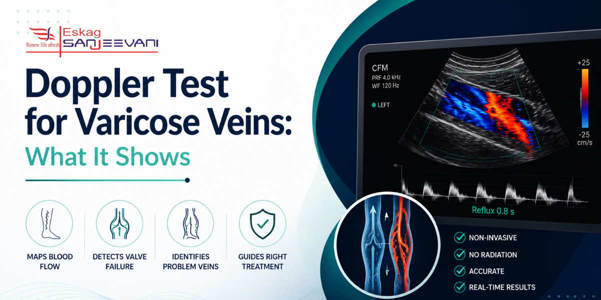
Varicose veins affect a significant proportion of Indian adults — yet most patients live with symptoms for years. A Doppler test for varicose veins maps blood flow through leg veins in real time. It identifies which valves are failing and provides evidence that determines whether treatment is needed. In a study at AIIMS Mangalagiri, Doppler ultrasound was performed in 70.6% of cases of varicose veins.
In this blog, we cover what the scan shows, how it is performed, and how results guide treatment.
Key Takeaways:
- Duplex Doppler ultrasound is the diagnostic test of choice for venous reflux; no varicose vein treatment should proceed without it.
- Reflux lasting more than 0.5 seconds on Doppler defines valve incompetence; this single threshold determines treatment eligibility.
- A Doppler scan for varicose veins also screens for DVT; concurrent DVT prevents ablation or sclerotherapy until resolved.
Quick Answer: A Doppler test for varicose veins maps venous reflux, identifies incompetent valves, assesses the saphenous system, and excludes DVT, taking 30-45 minutes with no radiation and no recovery time.
Quick Links
What Are Varicose Veins and When Should You Get a Doppler Scan
Varicose veins are not simply a cosmetic concern; they represent progressive venous valve failure, and a Doppler scan for varicose veins is the only investigation that maps the anatomy of that failure well enough to guide treatment safely.
- Varicose veins form when venous valves become incompetent, allowing blood to flow backwards (reflux) and pool in the superficial veins of the leg, causing them to dilate, elongate, and become visibly tortuous beneath the skin. The underlying problem is not the visible vein but the faulty valve above it, which only Doppler can locate.
- CEAP grades chronic venous disease from C0 (no visible disease) to C6 (active venous ulcer); the majority of Indian patients with varicose veins present at CEAP C2 or C3. In an Indian study of 150 patients with varicose veins, 38% presented with CEAP C3 (oedema) and 27.3% with CEAP C2 (visible varicose veins). Doppler scan is recommended from C2 onward when varicose veins become clinically visible and symptomatic.
- Duplex Doppler ultrasound is the diagnostic test of choice for evaluating venous reflux, recommended by the Society for Vascular Surgery, American Venous Forum, and American Vein and Lymphatic Society as the first-line investigation for all patients with symptomatic varicose veins before any treatment decision is made.
- Aching, heaviness, or swelling in the legs that worsens with prolonged standing; skin discolouration or thickening above the medial ankle; leg ulcers or healed ulcers; or varicose veins that cause documented cosmetic or functional concern at C2 or above are clinical indications for a Doppler scan of the legs before treatment is planned.
- In an Indian retrospective Doppler study of 200 patients with varicose veins at AIIMS Mangalagiri, 67% were male, and the left limb was most commonly affected [1]. Male patients aged 31-40 were the most prevalent group. In India, prolonged standing occupations, including teaching, surgery, factory work, and retail, are the most commonly associated risk factors for the development of varicose veins.
Now that you know when a Doppler scan is needed, here is exactly what the investigation reveals about your veins.
What Does a Doppler Ultrasound for Varicose Veins Actually Show?
A Doppler ultrasound for varicose veins is not simply a picture of the veins; it is a functional assessment of how blood moves through them, which valves are failing, and where the problem originates along the venous system.
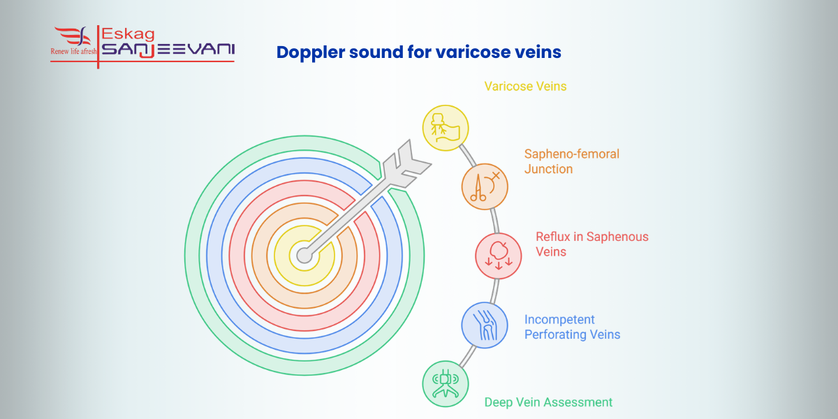
- Sapheno-femoral junction (SFJ): The SFJ is where the great saphenous vein drains into the deep femoral vein; incompetence here is the most common source of varicose vein reflux detected on Doppler ultrasound. This single finding directly determines whether the main trunk of the saphenous vein requires treatment.
- Great and small saphenous vein reflux: The scan measures GSV and SSV diameter and confirms whether valves are competent. A dilated GSV with confirmed reflux is the primary ablation or surgical target. Without Doppler, neither the extent nor the starting point of reflux can be established.
- Incompetent perforating veins: Perforators connect the superficial and deep venous systems; incompetent ones allow deep venous pressure to drive superficial reflux. Identifying and precisely mapping them on a Doppler scan of the legs is essential to prevent recurrence after treatment.
- Deep vein assessment (DVT) exclusion: The scan evaluates the deep veins to exclude DVT. A patient with concurrent DVT cannot safely receive varicose vein treatment until the clot is resolved; the assessment happens during the same appointment.
- Advanced disease findings: In patients with haemosiderin staining, lipodermatosclerosis, or active venous ulcers, a Doppler ultrasound for varicose veins maps the responsible incompetent veins, so the surgeon addresses the source of the disease, not just the visible surface abnormality.
With a clear picture of what the scan shows, here is exactly how the Doppler scan for varicose veins is performed.
5 Steps to Perform the Doppler Scan for Varicose Veins
A Doppler scan for varicose veins follows a structured, stepwise protocol. Understanding each step removes anxiety before the appointment and helps you know what to report to the sonographer during the scan.
Here is a step-by-step process to perform a Doppler scan for varicose veins:
Step 1: No Special Preparation Required
No fasting, no medication changes, and no contrast injection is needed for a venous Doppler scan. Wear loose, comfortable clothing that allows easy access to the full length of both legs, from the groin to the ankles. Remove nail polish if toe veins need assessment.
Step 2: Standing Position for Accurate Results
The scan is performed with you standing; gravity-dependent venous filling in the upright position is what makes reflux visible on the screen. Lying down misses most clinically significant venous incompetence, which is why patient positioning is protocol-critical rather than optional.
Step 3: Gel Application and Transducer Placement
A clear gel is applied to the skin from the groin to the ankle; this eliminates air between the skin and transducer and allows sound waves to transmit clearly. The sonographer moves the probe systematically along the great saphenous vein, small saphenous vein, perforators, and deep veins.
Step 4: Compression to Provoke Reflux
The sonographer performs intermittent calf and thigh compression; upon release, colour Doppler shows whether blood flows backwards through incompetent valves. Reflux lasting more than 0.5 seconds in the GSV or SSV confirms valve incompetence and defines the vein as treatment-eligible.
Step 5: Venous Mapping and Report
After identifying all incompetent veins and reflux patterns, the sonographer produces a venous map, a diagram of the full distribution of disease that the vascular surgeon uses for pre-operative planning.
Also read: How Abdominal Ultrasound Detects Early Abdominal Issues.
Doppler Scan for Varicose Veins vs DVT: Is It the Same Test?
A Doppler scan for varicose veins and a DVT scan use the same machine — but they answer entirely different clinical questions, follow different protocols, and have different consequences when findings are positive.
| Parameter | Varicose Vein Doppler Scan | DVT Doppler Scan |
| Patient position | Standing, gravity fills superficial veins to make reflux visible | Lying down, deep vein compression is assessed at rest |
| What it detects | Valve incompetence, venous reflux, saphenous vein involvement | Non-compressible deep vein clot, acute or chronic thrombosis |
| Compression technique | Calf compression provokes reflux through incompetent superficial valves | Deep vein compression confirms or excludes a clot by compressibility |
| Urgency of result | Guides elective treatment planning, not time-critical | Potentially urgent, undetected DVT can propagate to pulmonary embolism |
| When both are done together | Varicose vein scans routinely screen deep veins in the same appointment. DVT must be excluded before any varicose vein treatment proceeds | DVT scan may identify concurrent superficial venous disease, but is not a substitute for a full varicose vein reflux assessment |
How to Treat Varicose Veins After a Doppler Scan
The Doppler scan determines which treatment is appropriate, at which anatomical level, and in which sequence. The visible vein is never the target; the Doppler-confirmed reflux source is. For GSV reflux at the sapheno-femoral junction, endovenous laser ablation or radiofrequency ablation is now the preferred first-line treatment globally. Foam sclerotherapy addresses tributary varicosities and incompetent perforators identified on the venous map. For CEAP C2 patients without SFJ incompetence, graduated compression stockings remain the correct first step, and the Doppler scan is what establishes this without unnecessary intervention. For patients in Kolkata, the best USG centre in Kolkata for venous Doppler is one with dedicated vascular protocols. Eskag Sanjeevani reports are structured to provide your vascular surgeon with the anatomical precision treatment planning requires.
Final Thoughts
A Doppler test for varicose veins is the first step that makes every subsequent treatment decision possible. If you have visible varicose veins with aching, swelling, or skin changes, do not wait for symptoms to worsen. CEAP C2 and above is the clinical threshold for investigation; catching disease at C2 produces better outcomes than waiting until C5 or C6. Ask whether the sonographer will assess deep veins for DVT simultaneously; it is standard protocol. At Eskag Sanjeevani, venous Doppler scans follow dedicated vascular protocols that give your surgeon the anatomical precision treatment planning requires.

Microbiologist with over 2 years of experience in medical writing, specialising in evidence-based healthcare content.

Clinical experience in diagnosis, treatment, and evidence-based patient care across a range of conditions.
References
- Arumugam S, Manaswini DP, Reddy PA, Ghoshal JA, Subbiah NK. A Study of Valvular Incompetencies in the Lower Limb Veins Using Doppler Ultrasound Findings. Cureus. 2024 Feb 7;16(2):e53765.
Duplex Doppler ultrasound is the diagnostic test of choice for evaluating venous reflux; no varicose vein treatment should proceed without it. The scan identifies which valves are failing and whether the deep veins contain clots, both of which directly determine the appropriate treatment.
A Doppler scan for varicose veins is completely painless, with no needles, no radiation, and no contrast injection. Mild pressure from the transducer and pulsing sounds during the scan are both normal and expected.
A Doppler scan is not required for minor spider veins without symptoms, but it is mandatory before any treatment procedure or when skin changes, ulcers, pain, or swelling are present. Without Doppler, the source of reflux cannot be identified and treatment cannot be safely planned.
Reflux lasting more than 0.5 seconds in the great or small saphenous vein confirms valve incompetence, defining the vein as treatment-eligible. Reflux below this threshold is physiological and does not require intervention.
Yes, a complete venous Doppler examination assesses the superficial system for reflux and the deep veins for thrombosis in the same appointment. A patient with concurrent DVT cannot safely receive ablation or sclerotherapy until the clot is fully resolved.