

 Medically Reviewed
Colour Doppler Test: What It Shows About Your Heart
A colour Doppler test is a specialised ultrasound examination that adds real-time, colour-coded blood flow information to a standard cardiac image, showing not just the structure of your heart but also how blood actually moves through it. Unlike a standard ECG or chest X-ray, the echo colour Doppler captures functional abnormalities, valve leaks, septal defects, and pressure changes- that structural imaging alone cannot detect. The test is non-invasive, uses no radiation, and is the standard first-line cardiac evaluation at most diagnostic centres and heart hospitals across India.
In this blog, we cover how a colour Doppler test works, what the colours actually mean, how it compares to a 2D echo, what it detects about your heart, and when a more advanced investigation is needed.
Key Takeaways:
- A colour Doppler test shows blood flow direction and speed in real time: red indicates flow towards the probe, blue indicates flow away from the probe, and it is not dangerous.
- Echo colour Doppler and 2D echo are not the same test; colour Doppler is a functional layer added to the structural 2D image.
- Colour Doppler cannot visualise coronary arteries; CT angiography or cardiac MRI is the appropriate next step when coronary disease is suspected.
Quick Answer: A colour Doppler test uses sound waves to show blood flow direction and speed through the heart in real time, detecting valve disease, septal defects, ejection fraction, and diastolic dysfunction without radiation.
Quick Links

What Is a Colour Doppler Test and How Does It Work?
A colour Doppler test is a specialised ultrasound examination that adds real-time colour-coded blood flow information on top of a standard greyscale image — showing not just heart structure, but how blood moves through it at every moment of the cardiac cycle.
- How it works: High-frequency sound waves reflect off moving red blood cells; a computer converts the frequency shift between the emitted and reflected waves into the colour-coded flow map shown on screen.
- What the colours mean: The BART convention applies; Blue Away, Red Towards. Blood flowing towards the probe appears red; blood flowing away appears blue. Brighter shades mean faster flow. The colours indicate direction only, not oxygenation.
- What a mosaic pattern signals: When blood velocity exceeds the machine’s display scale, a mixed mosaic pattern appears, known as aliasing. This is routinely seen in valve stenosis and regurgitation and is a diagnostic cue rather than a visual error.
- No radiation involved: A colour Doppler test uses sound waves only, no ionising radiation, no contrast injection, and no recovery time. It is safe for all ages, including children and pregnant women.
- What it shows that greyscale ultrasound cannot: Standard ultrasound shows only cardiac structure. The colour Doppler layer detects valve leaks, septal defects, and abnormal flow jets that are entirely invisible on a structural image alone.
Colour Doppler vs 2D Echo: What Is the Actual Difference?
The terms “2D echo” and “echo colour Doppler” are used interchangeably across most Indian diagnostic referrals; they are not the same test, and understanding the difference helps you know exactly what your doctor has ordered and why.
Here is a tabular representation to understand the difference between the two:
| Parameter | 2D Echo | Echo Colour Doppler |
| What it shows | Heart structure, chambers, walls, and valves in black and white | Blood flow direction, velocity, and turbulence in real-time colour |
| What it misses | Functional abnormalities, a leaking valve, may appear normal on 2D | Cannot replace structural 2D assessment; both layers are needed |
| Clinical relationship | The structural foundation of every echocardiogram | An additional layer applied on top of 2D, not a separate test |
| When ordered alone | Rapid emergency assessment of wall motion or pericardial effusion | Never ordered alone, always combined with 2D |
| Combined study detects | Valve stenosis, regurgitation, septal defects, ejection fraction, diastolic dysfunction | The standard cardiac Doppler ultrasound evaluation is ordered across India |
Now, let’s explore how colour Doppler tests actually work and what they tell us about the heart.
What Does a Colour Doppler Test Show About the Heart?
A colour Doppler test for heart assessment goes beyond structure; it shows whether blood moves correctly through each chamber and valve, and quantifies the severity of any abnormality well before a patient becomes symptomatic.
- Valve stenosis and regurgitation: Colour Doppler identifies narrowed valves (stenosis) and leaking valves (regurgitation—backflow jets in the wrong direction). Regurgitation is graded from trace to severe. Trace regurgitation is present in up to 70% of healthy adults and requires no treatment.
- Ejection fraction: Normal EF range is 55-70%; EF below 40% indicates heart failure with reduced ejection fraction. In an Indian study of 75 HFrEF patients at Madras Medical College (2024-25), pulmonary hypertension and right ventricular dysfunction independently predicted adverse outcomes, findings made possible by colour Doppler assessment [1].
- Diastolic function: Approximately 30-40% of heart failure patients have normal EF but abnormal diastolic relaxation, detectable only by Doppler echocardiography. This pattern is common in Indian hypertensive patients and frequently precedes symptoms by years.
- Congenital defects: Septal defects are detected as abnormal colour jets crossing the septum. India has an estimated 200,000 new congenital heart disease births annually; colour Doppler is the primary diagnostic tool for these defects across Indian cardiac centres.
- Pulmonary artery pressure: Colour Doppler estimates pulmonary artery systolic pressure non-invasively; elevated pressures indicating pulmonary hypertension can be identified without cardiac catheterisation. Pulmonary hypertension independently predicted higher in-hospital mortality.
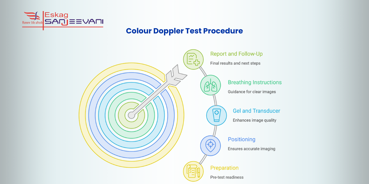
The Colour Doppler Test Procedure: What to Expect
A colour Doppler test for the heart is a non-invasive, painless procedure that takes 30-60 minutes. Knowing what happens at each step removes the uncertainty that causes most patient anxiety before the appointment.
Here is a stepwise process for the colour doppler test and what to expect from the test:
Step 1: Preparation
No fasting is required for a standard cardiac colour Doppler; you may eat, drink, and take medications as normal. Wear loose, comfortable clothing, remove jewellery, and bring all previous echo or cardiac reports for direct comparison during the examination.
Step 2: Positioning
You will be asked to lie on your left side on the examination table; this brings the heart closer to the anterior chest wall, improving acoustic access and image quality. This positioning is standard and is why the technician will ask you to turn partway through the scan.
Step 3: Gel and Transducer
The technician applies a colourless gel to your chest to eliminate air between the skin and the transducer; air blocks sound waves, degrading image quality. The transducer is then moved to four standard positions on the chest to capture all required cardiac views.
Step 4: Breathing Instructions
You will be asked to hold your breath briefly at specific moments and to breathe deeply or inhale through your nose occasionally. This temporarily removes lung interference from the image; it is standard practice and not a sign of anything unexpected.
Step 5: Report and Follow-Up
Once imaging is complete, the gel is wiped off, and you can resume all normal activities immediately; there is no recovery time. At diagnostic centres, reports are available the same day or within 24 hours and should be reviewed by a cardiologist for any complex findings.
When Colour Doppler Is Not Enough: What Comes Next
A colour Doppler test is the correct first-line cardiac investigation for most patients, but several clinical scenarios fall outside its diagnostic scope, and knowing when to request the next step prevents diagnostic delays.
- Poor acoustic windows: Variable image quality and limited acoustic windows are documented limitations of echocardiography, affecting patients with obesity, chest deformity, or COPD. In severe cases, cardiac MRI is recommended instead, providing complete cardiac assessment without acoustic window restrictions.
- Coronary artery disease: Colour Doppler cannot visualise coronary arteries, which measure 2-4 mm and move rapidly with each heartbeat, beyond the echo’s spatial resolution. CT coronary angiography is the non-invasive test of choice when coronary disease is suspected.
- Myocardial tissue characterisation: Cardiac MRI is the gold standard for myocardial scar quantification, cardiomyopathy characterisation, and pericardial disease, findings that colour Doppler can suggest but cannot definitively characterise.
- Carotid and stroke assessment: In an Indian multicentre study, CT angiography and MR angiography identified 40 and 43 vulnerable carotid plaques, respectively [2], whereas colour Doppler lacked sensitivity for detecting vulnerable plaques. CTA and MRA are now preferred in acute stroke scenarios.
Final Thoughts
A colour Doppler test for heart assessment is one of the most clinically complete non-invasive investigations available, and for most Indian patients referred for cardiac symptoms, it is the correct and sufficient starting point. Understanding what the colours mean, what each section of your report describes, and when the test has reached its diagnostic limits makes you a more informed participant in your own care. If your report says “limited study” or “recommend further evaluation,” do not wait; ask your cardiologist directly whether cardiac MRI or CT angiography is the next clinically indicated step. An echo colour Doppler result is not a final verdict; it is a detailed clinical snapshot that your cardiologist interprets alongside your symptoms, history, and other investigations. At Eskag Sanjeevani Hospitals, cardiac colour Doppler examinations are reported by cardiologist-led teams, ensuring the findings you receive are interpreted with the clinical precision that cardiac diagnosis requires.

Microbiologist with over 2 years of experience in medical writing, specialising in evidence-based healthcare content.

Clinical experience in diagnosis, treatment, and evidence-based patient care across a range of conditions.
References
- Irfan S, Padmakaran V, Prasanth R, Nazar S, Srinivas S, Tv AK, Bhandary VS, Patel PV, Baldota R, Puthenveedu Soman A, Vattapothy BA. Clinical and Echocardiographic Profile of Patients With Heart Failure With Reduced Ejection Fraction: A Retrospective Outcome Analysis. Cureus. 2025 Oct 24;17(10):e95354.
- Saluja KV, Swami MK, Pillai D, Meena M, Meena DR. Getting to the heart of Carotid and Vertebral imaging in acute ischemic stroke: an all-encompassing cross-sectional comparative analysis of Colour Doppler Ultrasound, CT Angiography, and MR Angiography. BJR Open. 2025 Dec 17;8(1):tzaf031.
A colour Doppler test is a specialised ultrasound examination that shows both heart structure and real-time blood flow direction and speed, using colour to map how blood moves through chambers, valves, and vessels. It detects valve stenosis and regurgitation, ejection fraction, congenital defects, diastolic dysfunction, and pulmonary artery pressure without radiation.
The colours indicate direction only, not oxygenation, danger, or severity. The BART convention applies: blood flowing towards the probe appears red, blood flowing away appears blue. Brighter shades mean faster flow; a mosaic or multicolour pattern indicates turbulent flow and is a diagnostic cue for the cardiologist, not a cause for patient concern.
A 2D echo shows heart structure in black and white, chamber sizes, wall thickness, and valve morphology. An echo colour Doppler adds a functional layer on top, showing blood flow direction and velocity in real time. The two are complementary, not competing, and most cardiac referrals in India order both together as a standard combined study.
A colour Doppler test is completely non-invasive and painless. It uses no ionising radiation, making it safe for patients of all ages, including children, pregnant women, and those requiring repeated cardiac follow-up. The only sensations are mild pressure from the transducer and the cool gel applied to the chest.
Colour Doppler cannot visualise the coronary arteries and has reduced accuracy in patients with poor acoustic windows; conditions like obesity or COPD can limit image quality. CT coronary angiography is preferred for suspected coronary artery disease, while cardiac MRI is recommended for myocardial scar quantification, cardiomyopathy characterisation, and cases where the echo window is insufficient.