
How Abdominal Ultrasound Detects Early Abdominal Issues
Many abdominal conditions develop gradually, often without causing obvious symptoms in their early stages. A person may experience occasional discomfort, mild bloating, or changes in digestion without realising that an underlying issue is affecting various. This may include organs such as the liver, gallbladder, kidneys, pancreas, or spleen. This is where an abdominal ultrasound becomes an important diagnostic tool. By using sound waves to generate real-time images of internal structures, the examination helps doctors identify abnormalities before they progress into more serious health concerns [1]. As one of the most commonly performed imaging investigations, abdominal ultrasound continues to play a key role in the early detection and evaluation of various abdominal issues.
Key Takeaways
- An abdominal ultrasound can identify complex abnormalities in abdominal organs before symptoms become severe or concerning.
- The examination provides real-time imaging, making it suitable for diagnosis and follow-up check-ups.
- An early ultrasound test can help doctors understand abdominal symptoms thoroughly, guide treatment decisions, and monitor existing conditions effectively.
Quick Answer: An abdominal ultrasound is needed to detect and evaluate abdominal issues by creating detailed images of the abdomen’s internal organs.
What Is an Abdominal Ultrasound?
An abdominal ultrasound, often called a USG (Ultrasonography) scan, is a non-invasive imaging procedure that uses high-frequency sound waves to examine organs and structures inside the abdomen. Unlike other imaging techniques that rely on radiation, ultrasound creates images through reflected sound waves, making it a safe and widely used diagnostic investigation.
During the procedure, a transducer is moved over the skin over the stomach. The device sends sound waves into the body and receives returning echoes from internal organs. These echoes are converted into images that appear on a monitor, allowing doctors and other medical experts to assess organ size, shape, structure, and visible abnormalities in real time.
Advancements in USG have significantly improved image quality and diagnostic accuracy. Today, abdominal ultrasound is more often used as a first-line evaluation. It is easily accessible, affordable, efficient, and capable of evaluating multiple organs during a single examination.
Organs Commonly Evaluated During an Abdominal Ultrasound
| Organ | Common Findings Assessed |
| Liver | Fatty liver disease, enlargement, cysts, masses |
| Gallbladder | Gallstones, inflammation, obstruction |
| Kidneys | Stones, cysts, swelling, structural abnormalities |
| Pancreas | Inflammation and visible structural changes |
| Spleen | Enlargement and abnormalities |
| Urinary Bladder | Stones, retention, wall irregularities |
| Blood Vessels | Blood flow and vascular abnormalities |
In certain situations, doctors may recommend an abdominal and pelvic ultrasound to provide a broader assessment of both abdominal and pelvic structures.
What Can an Abdominal Ultrasound Detect?
One of the greatest strengths of abdominal ultrasound is its ability to identify abnormalities that may not be apparent during a routine physical examination. The test provides valuable information about multiple organs (of the stomach) and often serves as the starting point for further medical evaluation.
Some of the abnormalities that may be identified include:
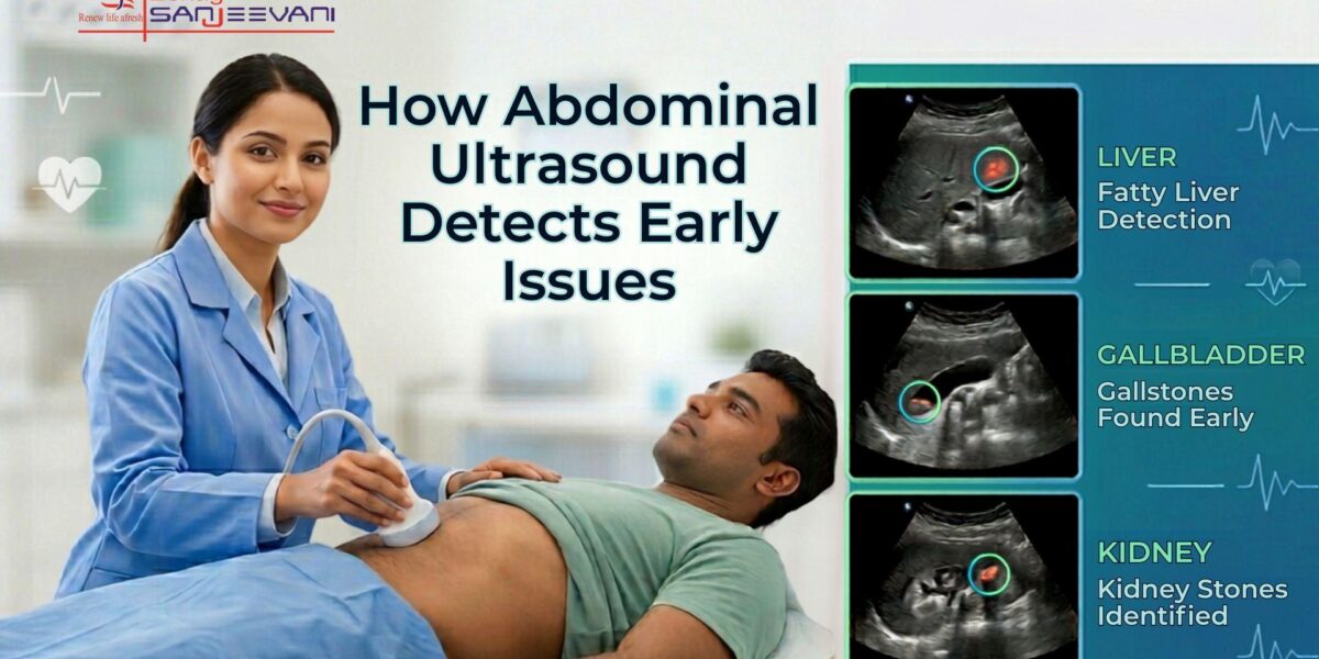
- Gallstones can often be visualised clearly on ultrasound, helping explain recurrent abdominal discomfort, nausea, or digestive symptoms after meals [1].
- Kidney stones, urinary tract obstruction, and swelling within the kidneys may be detected before they lead to significant complications [1].
- Fatty liver disease and liver enlargement can frequently be identified during routine abdominal ultrasound examinations, even in asymptomatic individuals.
- Fluid accumulation within the abdominal cavity may indicate underlying medical conditions requiring further investigation and clinical management.
- Certain cysts, benign growths, and structural abnormalities affecting abdominal organs can be recognised during detailed ultrasound assessment.
The ability to evaluate multiple organs simultaneously makes abdominal ultrasound a valuable diagnostic tool for investigating both specific symptoms and broader abdominal concerns.
Also Read: USG Scan in Pregnancy: A Simple Guide for Indian Parents
Why Is an Abdominal Ultrasound Recommended?
Doctors recommend abdominal ultrasound for a variety of reasons. In many cases, symptoms alone may not provide enough information to determine the underlying cause of a patient’s condition. Imaging allows doctors to examine internal organs directly and identify abnormalities that may require treatment or monitoring.
The following are situations in which a USG may be recommended:
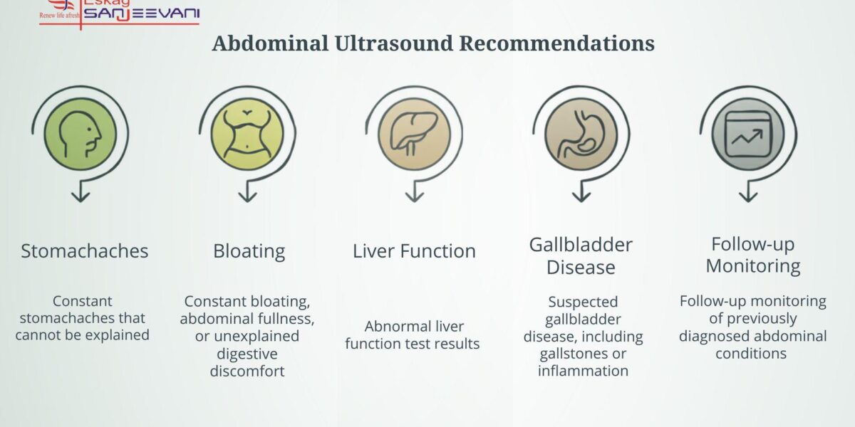
- Constant stomachaches that cannot be explained by physical examination often need imaging for further evaluation.
- Constant bloating, abdominal fullness, or unexplained digestive discomfort may indicate underlying abnormalities involving abdominal organs.
- Abnormal liver function test results frequently prompt doctors to recommend abdominal ultrasound for a detailed assessment of liver health.
- Suspected gallbladder disease, including gallstones or inflammation, can often be evaluated effectively through USG.
- Follow-up monitoring of previously diagnosed abdominal conditions may require USG to assess recent changes.
Identifying abnormalities at an earlier stage can help doctors make informed decisions about additional treatment plans or long-term monitoring strategies.
Preparing for an Abdominal Ultrasound: What to Expect
No matter what the illness is, a doctor’s visit, a simple test, or an USG is concerning for many patients. Thus, understanding the process beforehand helps reduce anxiety and ensures smoother preparation. The exact instructions may vary depending on the organs being assessed.
Step 1: Patients are often advised to avoid food for several hours before the procedure. Fasting can help improve visualisation of certain abdominal structures, particularly the gallbladder. In some situations, particularly when pelvic structures are also being examined, patients may be asked to drink water beforehand to ensure adequate bladder filling.
Step 2: The procedure is simple. A clear gel is applied to the skin, and the sonographer moves the transducer over the abdomen while capturing images from multiple angles. The examination is typically painless and usually completed within 20 to 40 minutes [2].
Step 3: Following the scan, patients can generally return to their normal activities immediately. The images are reviewed by a radiologist, and the findings are shared with the referring doctor, who interprets the results within the broader clinical context.
Why Ultrasound Remains Important for Early Detection
Despite the availability of advanced imaging technologies, abdominal ultrasound continues to play an important role in diagnosis. It provides live visualisation of internal organs, combined with its safety profile and accessibility. This is why it is often the first test recommended for many abdominal issues.
Unlike other tests that involve radiation exposure, ultrasound can be repeated as needed, making it extremely useful for monitoring chronic conditions. It also allows doctors to efficiently assess organ conditions, often providing answers that guide subsequent diagnostic or therapeutic decisions [2].
This growth of ultrasound scanning has further expanded the range of conditions that can be evaluated. As a result, it remains one of the most widely trusted tools for detecting abdominal problems at an earlier stage.
Why Choose Eskag Sanjeevani for Abdominal Ultrasounds?
At Eskag Sanjeevani, we provide the best to all our patients, from imaging services and support and follow-up check-ups to accurate diagnosis and coordinated care. Diagnostic accuracy depends not only on technology and infrastructure but also on the dedication and experience of the professionals performing and examining the patients.
Eskag Sanjeevani is one of the leading hospitals in Kolkata, combining advanced infrastructure with experienced radiologists and trained medical teams who understand the importance of detailed, reliable imaging assessments. Whether the requirement is a routine abdominal ultrasound, an ultrasound abdomen and pelvis examination, or a specialised USG in a hospital setting, we want you to benefit from our services.
For individuals seeking the best USG centre in Kolkata, access to high-quality imaging technology, medical expertise, and best healthcare services can significantly support timely diagnosis and informed treatment planning. Along with dedicated medical experts, we provide affordable treatments and facilities, as we understand that every patient’s primary concern is cost.
Wrapping Up
Many abdominal problems begin with minor changes that remain unnoticed until symptoms become more prominent. An early abdominal ultrasound helps detect problems before they worsen by providing a detailed view of internal organs and surrounding structures without the need for invasive or complex procedures.
From evaluating abdominal pain and digestive symptoms to identifying gallstones, kidney abnormalities, liver conditions, and other abdominal issues, ultrasound remains one of the most valuable diagnostic tools. When used appropriately and interpreted within the broader clinical picture, it can provide crucial information that supports early detection, accurate diagnosis, and effective patient management. Stop waiting for the symptoms to worsen and book your appointment now.


References
- Kurzweil, Ami, and Jennifer Martin. “Transabdominal Ultrasound.” PubMed, StatPearls Publishing, 2020.
- Cleveland Clinic. “Abdominal Ultrasound: What It Is, Types, Details.” Cleveland Clinic, 2023.
An abdominal ultrasound can detect gallstones, kidney stones, fatty liver disease, cysts, fluid accumulation, organ enlargement, and several other abdominal abnormalities.
Yes. Abdominal ultrasound uses sound waves rather than radiation, making it a safe and commonly performed imaging procedure.
You might be advised to avoid eating or drinking for several hours before a USG to improve image quality. Depending on the area being examined, you may also be asked to drink water and arrive with a full bladder. Most examinations take approximately 20 to 40 minutes and are a simple procedure.
Many abdominal ultrasound examinations require fasting for several hours, although instructions may vary. You may drink water but avoid drinking milk or juice.
An abdominal ultrasound may be recommended, as it focuses on the abdominal organs and helps detect unexplained symptoms and underlying conditions, so doctors can provide a treatment plan that best suits your condition.