

 Medically Reviewed
Sinus CT Scan: Why Chronic Headaches Need Imaging
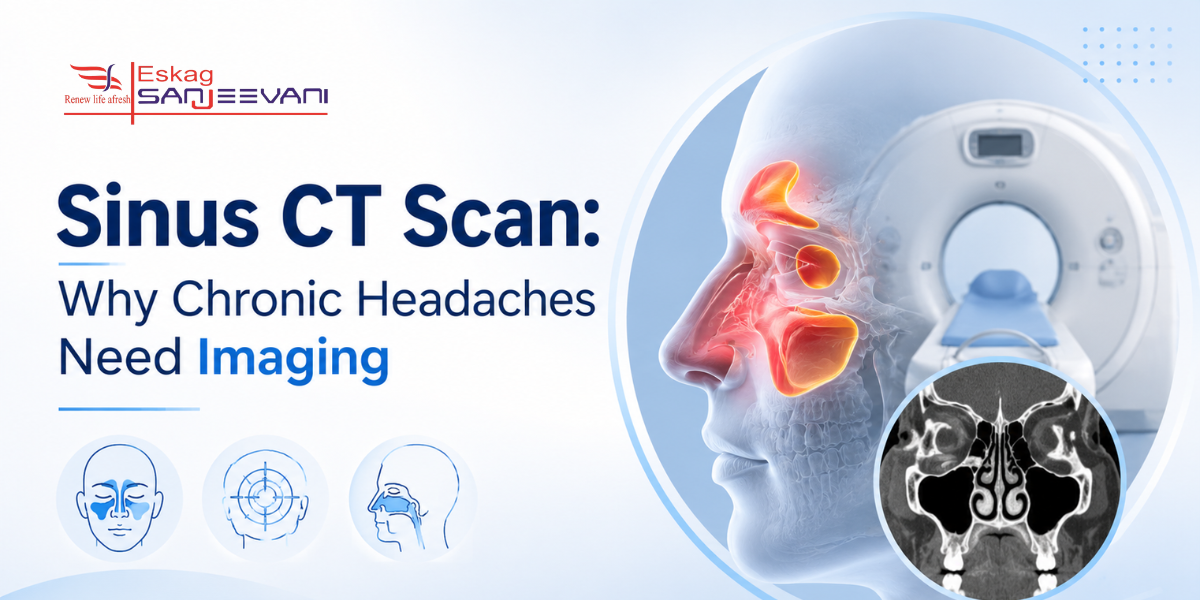
Living with persistent headaches, facial pressure, and blocked sinuses is exhausting, and arriving at the right diagnosis takes longer than it should for most Indian patients. A sinus CT scan is the gold-standard imaging tool for evaluating the paranasal sinuses, providing detailed information on bony anatomy and mucosal disease that clinical examination and plain X-ray simply cannot offer. What makes it clinically essential is not just what it detects, but what it rules out, because the majority of patients who believe they have a sinus headache are eventually diagnosed with migraine, and a normal sinus CT is what redirects them to the right care. In India, where allergic rhinosinusitis affects 20-30% of urban populations and chronic headaches are among the most common ENT and neurology referrals, the decision of when to order a sinus CT and how to interpret it carries real clinical weight.
In this blog, we cover when to do a sinus CT scan, what it shows, how to interpret a normal result, how it differentiates sinus headache from migraine, and how it compares with MRI.
Key Takeaways:
- A sinus CT scan takes place only after 12 weeks of symptoms despite treatment, not for a single acute episode.
- 74.3% of “sinus headache” patients in a prospective study met IHS migraine criteria, and CT ruled out sinusitis in most.
- A normal sinus CT does not mean symptoms are imaginary; it redirects the diagnosis toward migraine, rhinitis, or nerve pain.
Quick Answer: A sinus CT scan images the paranasal sinuses in cross-section to detect mucosal thickening, opacification, polyps, and anatomical variants, and to differentiate true sinusitis from migraine or other headache causes.
Quick Links
What Is a Sinus CT Scan and When Is It Ordered?
A sinus CT scan, also called a CT of the sinuses or a CT scan of the paranasal sinuses, is the gold-standard imaging tool for evaluating sinus anatomy and disease, providing detailed imaging of bony structures and mucosal changes that clinical examination and plain X-ray cannot offer.
- The paranasal sinuses are four paired air-filled spaces surrounding the nasal cavity: the maxillary, ethmoid, frontal, and sphenoid sinuses. A CT of the sinuses captures all four sinus groups simultaneously in cross-section, along with the nasal cavity, ostiomeatal complex, and surrounding bony architecture.
- A CT scan for sinusitis is the imaging of choice for patients with recurrent acute or chronic rhinosinusitis, defined as sinus inflammation lasting more than 12 weeks despite treatment, per the AAO-HNSF 2025 updated guideline. It is also ordered before functional endoscopic sinus surgery, when complications are suspected, and when initial medical treatment has failed.
- A CT scan for sinusitis is not recommended at first presentation of uncomplicated acute rhinosinusitis; routine CT without red flag features exposes patients to unnecessary radiation and rarely changes early management decisions.
- A dedicated CT of the sinuses delivers approximately 0.5-1.0 mSv, significantly less than a standard CT head. Low-dose protocols now available at most Indian CT scan hospitals reduce this further, making repeat imaging appropriate for recurrent disease monitoring.
- Among 113 patients undergoing CT scans for sinusitis at a tertiary care centre, 96.46% had sinusitis; mucosal thickening was the most common finding in both acute and chronic cases [1]. In India, where air pollution exacerbates mucosal inflammation, sinus CT referrals are increasing at ENT centres in cities including Kolkata.
What Does a Sinus CT Scan Actually Show?
A CT scan for sinusitis shows five clinically actionable findings: mucosal thickening, sinus opacification (fluid or pus filling the sinus cavity), nasal polyps, anatomical variants such as a deviated septum or concha bullosa, and bony changes including erosion or sclerosis. The ostiomeatal complex, the drainage pathway of the anterior sinuses, is the most critical structure a CT of the sinuses visualises, as its blockage is the root cause of most chronic sinusitis. Radiologists grade severity using the Lund-Mackay scoring system; each sinus group is scored from 0 to 2, for a maximum total of 24. A score above 4 is generally considered clinically significant and directly guides surgical planning.
Normal Sinus CT but Symptoms Persist: What It Means
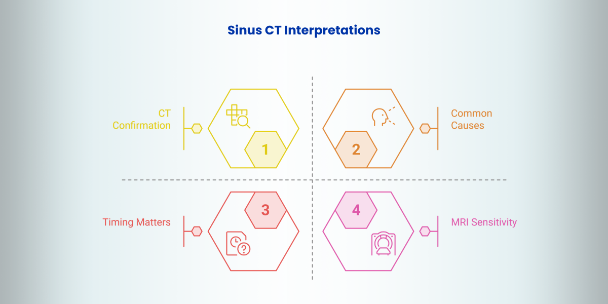
A normal sinus CT report does not mean your symptoms are imaginary — it means structural sinus blockage is not the cause, which is clinically useful information that redirects the diagnostic pathway rather than ending it.
- What a normal CT actually confirms: A CT scan of the sinuses is excellent at identifying physical obstruction, mucosal thickening, polyps, fluid, and bony abnormalities. When these are absent, an active sinus infection or significant chronic sinusitis is an unlikely cause. The scan does not assess neurological conditions, allergic inflammation without blockage, or nerve sensitivity.
- Common causes of sinus-like symptoms with a normal CT: Migraine is the most frequent missed diagnosis; it causes facial pressure, nasal congestion, and periorbital pain without any CT findings because it is neurological, not structural. Allergic rhinitis, non-allergic rhinitis, temporomandibular joint dysfunction, and silent reflux all produce sinus-like symptoms with a clear CT scan.
- Timing affects the result: CT is a snapshot of one day; someone scanned between flares may show a normal result even when significant disease is present during episodes. Recurrent acute sinusitis, by definition, has symptom-free intervals during which CT may appear entirely clear.
- MRI is more sensitive than CT for mucosal disease: A PMC study confirmed a significant correlation between MRI sinus findings and patient symptomatology, with no correlation in the CT group, indicating that symptomatic patients with normal CT may have findings detectable only on MRI.
Now, let’s understand the difference between migraine and sinus headache.
Sinus Headache vs Migraine: What a CT Scan Shows
One of the most clinically useful things a sinus CT scan reveals is not what it shows, but what it rules out, which is precisely what allows an ENT to separate a genuine sinus headache from the far more common diagnosis of migraine.
Here is a tabular representation to understand the difference between the two:
| Parameter | Sinus Headache (True Sinusitis) | Migraine Misdiagnosed as Sinus |
| CT scan findings | Mucosal thickening above 6 mm, opacification, OMC blockage, fluid levels, all visible on CT | Normal or minor incidental findings; CT shows no significant sinus disease |
| Lund-Mackay score | A score above 4 indicates clinically significant disease | Score typically below 3, within the range seen in asymptomatic adults |
| Headache character | Dull pressure localised to the sinus region, worsens with bending forward, associated with coloured nasal discharge and fever | Throbbing, unilateral, worsens with physical activity, associated with nausea, light and sound sensitivity |
| Prevalence among “sinus headache” patients | Minority, in a prospective study of 35 “sinus headache” patients, 74.3% met IHS criteria for migraine, not sinusitis | The majority of studies show that up to 88% of self-diagnosed sinus headaches meet migraine diagnostic criteria |
| CT’s role in diagnosis | Confirms sinusitis and guides treatment, medical or surgical | Rules out sinusitis, redirects patient to neurology for migraine management |
| India-specific context | In an Indian study of 500 headache patients, sinusitis was found in only 11.6%; the large majority had no structural sinus pathology on imaging [2] | Migraine is significantly underdiagnosed in India; many patients receive repeated ENT referrals and antibiotic courses for what is clinically migraine |
Also read: Diagnosis and Relief: Ear Pain in Children During the Cold.
Sinus CT Scan vs MRI: Which One Does Your ENT Order?
A sinus CT scan and MRI are complementary; CT is the gold standard for anatomy and surgical planning, while MRI adds value when soft tissue characterisation or suspected complications go beyond what bony detail alone can answer.
| Parameter | Sinus CT Scan | MRI Paranasal Sinuses |
| Best for | Bony anatomy, sinus drainage pathways, anatomical variants, pre-FESS surgical planning, gold standard per AAO-HNSF 2025 | Soft tissue characterisation — differentiates tumour from inflammation, detects intracranial or orbital extension |
| When ordered | Chronic sinusitis, recurrent acute sinusitis, failed medical treatment, pre-surgical planning | Suspected sinonasal tumour, ambiguous CT findings, or when complications require soft tissue detail |
| Radiation | Low dose, 0.5-1.0 mSv; significantly less than standard CT head | No radiation, preferred for younger patients needing repeat imaging |
| When both are needed | CT maps bony anatomy for surgery | MRI assesses soft tissue when neoplasm or intracranial extension is clinically suspected, alongside CT findings |
Final Thoughts
A sinus CT scan is most useful when it is ordered for the right clinical reason at the right stage of illness, not as a first response to any headache, and not after years of untreated symptoms. If your scan shows significant disease, the Lund-Mackay score your radiologist assigns directly guides whether medical treatment or surgical referral is appropriate next. If your sinus CT scan comes back normal but symptoms persist, do not stop investigating; ask your ENT specifically whether migraine, allergic rhinitis, or nasal endoscopy should be the next step. Carrying your previous imaging on CD or digital format to every follow-up appointment allows direct comparison that changes clinical decisions more than a repeat examination alone.
At Eskag Sanjeevani, sinus CT reports are interpreted by subspecialist radiologists who communicate findings directly to your ENT team, ensuring that what the scan shows is translated accurately into what happens next in your care.

Microbiologist with over 2 years of experience in medical writing, specialising in evidence-based healthcare content.

Clinical experience in diagnosis, treatment, and evidence-based patient care across a range of conditions.
References
- Tuladhar AS, Bhattarai A, Bimali S, Pokharel B, Pradhan S, Shrestha A, Acharya R. Sinusitis among Patients Undergoing CT Scan of Paranasal Sinuses in a Tertiary Care Centre: A Descriptive Cross-sectional Study. JNMA J Nepal Med Assoc. 2022 Oct 1;60(254):857-860.
- Rai GS, Rai T, Jain L, Vyas MM, Roshan R. Evaluation of CT and MRI Findings among Patients Presented with Chief Complaint of Headache in Central India. J Clin Diagn Res. 2016 Feb;10(2):TC21-5.
A sinus CT scan is not recommended for uncomplicated acute sinusitis at first presentation; it is ordered when symptoms persist beyond 12 weeks despite treatment, when recurrent acute sinusitis occurs three or more times a year, or when functional endoscopic sinus surgery is being planned. Red-flag features such as eye swelling, vision changes, or severe worsening of headache warrant urgent imaging regardless of symptom duration.
A normal sinus CT scan is not a dead end; it provides valuable information by ruling out active sinus infection, narrowing the list of possible causes, and redirecting care toward more appropriate evaluation. Common causes of persistent sinus-like symptoms with a normal CT include migraine, allergic rhinitis, and nerve pain, all of which require a different clinical pathway from sinusitis treatment.
Doctors cannot diagnose migraines with a CT scan; the test helps rule out other causes of pain, such as sinus disease. In a prospective study of 35 patients presenting with “sinus headache,” 74.3% met IHS criteria for migraine. A normal sinus CT is what redirects these patients from ENT to neurology for the correct diagnosis.
A dedicated sinus CT delivers approximately 0.5-1.0 mSv, significantly less than a standard CT head and well within safe clinical limits. Low-dose protocols now available at most Indian CT scan hospitals further reduce this, making repeat imaging appropriate for recurrent disease monitoring without significant cumulative radiation concern.
CT is the gold standard for sinus imaging; it provides superior visualisation of bony anatomy, drainage pathways, and anatomical variants and is the mandatory pre-surgical tool for FESS planning. MRI is typically best for soft-tissue concerns and suspected complications, including suspected sinonasal tumour, intracranial extension of sinusitis, and cases where CT findings are ambiguous and soft-tissue detail changes management.