

 Medically Reviewed
Fracture vs Sprain: How an X-Ray Tells the Difference
A twisted ankle does not always mean a sprain. In a North Indian study of 140 patients with acute ankle or midfoot injuries, 48 patients had fractures, while the Ottawa Ankle Rules identified all fracture cases, showing 100% sensitivity and the potential to reduce unnecessary X-rays by about 51% [1]. Pain, swelling and bruising can occur with both a fracture and a sprain, making them difficult to distinguish without an examination.
This article explains the difference between a fracture and sprain, when an ankle or foot X-ray is appropriate, and how doctors decide what treatment you may need.
Key Takeaways
- A fracture breaks a bone, while a sprain injures a ligament. Both can cause pain, swelling, bruising and difficulty walking.
- X-rays can identify many bone fractures but do not directly show most ligament injuries. A normal X-ray therefore does not rule out a significant sprain or every type of bone injury.
- Fracture pain is often concentrated over the injured bone, while sprain pain is usually around the affected ligament and joint. However, pain patterns can overlap, so symptoms alone cannot confirm the injury.
Quick answer: A fracture is a break in the bone, while a sprain is a ligament injury; an X-ray is used when examination findings suggest a possible fracture.
- Fracture vs Sprain: What Is the Difference?
- Break or Sprain: What Symptoms Should You Check?
- What Does Indian Evidence Say About Ankle X-Rays?
- Is It Necessary to Get an X-Ray for an Ankle Sprain?
- When Is a Foot X-Ray Needed as per Ottawa Foot Rules?
- What Happens During an Ankle or Foot X-Ray?
- What If the X-Ray Shows a Fracture?
- What If the X-Ray Is Normal?
- Digital X-Ray Services at Eskag Sanjeevani Hospitals
Fracture vs Sprain: What Is the Difference?
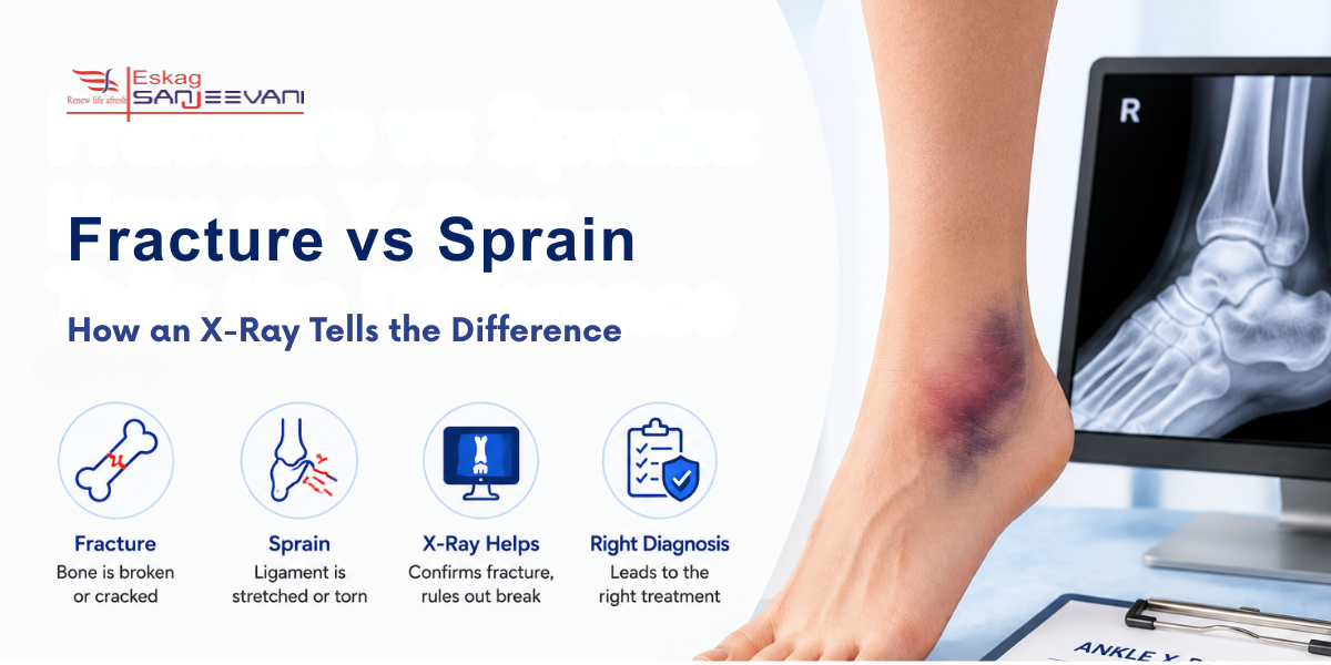
A fracture is a crack or break in a bone. A sprain is an injury to a ligament, the strong tissue connecting bones around a joint. Both commonly occur after a fall, sudden twist, sports injury or road traffic accident.
The following table briefly helps to learn the difference between a fracture and sprain.
| Feature | Fracture | Sprain |
| Main structure injured | Bone | Ligament |
| Common cause | Impact, fall or twisting force | Twisting, rolling or sudden movement |
| Pain | Often concentrated over a bone | Often around the joint or ligament |
| Swelling | Common | Common |
| Bruising | Can occur | Common |
| Walking | May be difficult or impossible | May be painful but possible |
| X-ray | Can show many fractures | Usually does not show ligament damage |
| Further imaging | CT or MRI in selected cases | MRI in selected persistent or complex injuries |
Break or Sprain: What Symptoms Should You Check?
The symptoms of ankle fracture vs sprain often overlap. Doctors therefore assess where the tenderness is located, how the injury occurred and whether you can bear weight.
Findings That Need Assessment for Fracture
- Tenderness directly over the ankle bones: Tenderness along the posterior edge or tip of the medial or lateral malleolus is an important criterion for considering an X-ray. The Ottawa Ankle Rules showed a pooled sensitivity of 4% for ankle fractures across 66 studies [2].
- Inability to take four steps: Inability to bear weight immediately after the injury and during clinical assessment increases the need for ankle or foot radiography.
- Severe pain after significant trauma: A high-force injury requires careful assessment because clinical symptoms alone cannot reliably exclude a fracture.
- Visible deformity: An abnormal ankle position or obvious change in alignment requires urgent assessment for a displaced fracture.
- Open wound near the injured bone: An open injury with suspected bone exposure can indicate an open fracture and requires urgent treatment.
- Numbness or reduced sensation: Altered sensation can indicate associated nerve injury and requires prompt examination.
- Pale, blue or unusually cold toes: These changes can indicate impaired blood flow and require urgent medical assessment.
Findings That Can Occur With a Sprain
Pain, tenderness, swelling and bruising are common after an acute ankle sprain. Clinical examination typically assesses swelling, bruising, tenderness and ligament stability to determine the extent of injury.
- Soft-tissue tenderness: Tenderness around the ankle ligaments, rather than directly over the bony landmarks, supports a ligament injury.
- Swelling: Swelling around the ankle is common after a sprain and can develop soon after the injury because of soft-tissue damage and inflammation.
- Bruising: Discolouration can occur as damaged small blood vessels bleed into the surrounding tissues.
- Pain with movement or weight-bearing: A sprain can make walking and ankle movement painful, particularly during the first few days.
- No obvious deformity: Most sprains do not cause visible changes in bone alignment.
What Does Indian Evidence Say About Ankle X-Rays?
The Ottawa Ankle Rules have also been studied in an Indian clinical setting. A prospective study from a teaching hospital in North India assessed 140 patients with acute ankle or midfoot injuries [1]. The rules showed:
- 100% sensitivity for detecting fractures
- 100% negative predictive value
- 7% specificity
- Potential reduction of radiographs by approximately 51%
Among the fractures identified in the study, 70.83% involved the malleolar zone, while 29.16% involved the midfoot. Fractures of the lateral malleolus accounted for 43.75% of all fractures.
Is It Necessary to Get an X-Ray for an Ankle Sprain?
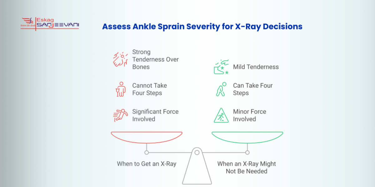
Primarily, no. An X-ray is not required for every ankle sprain. Doctors first examine where the pain is located, check for tenderness over the bones and assess whether you can walk. An ankle X-ray is more likely to be recommended when:
- There is strong tenderness over the ankle bones: Pain directly over the hard bony areas on either side of the ankle can indicate a possible fracture.
- You cannot take four steps: Difficulty bearing weight both immediately after the injury and during the examination increases the need for an X-ray.
- The injury involved significant force: A major fall, road accident, or strong impact requires a careful assessment for possible bone injury.
- The ankle looks deformed: An unusual shape or position often indicates a displaced fracture and requires prompt medical attention.
- The injury cannot be assessed reliably: Severe swelling, pain or other factors can make the examination difficult.
- Pain or swelling is unusually severe or does not improve: Prolonged or worsening symptoms after an initial assessment can require repeat examination or further imaging.
When Is a Foot X-Ray Needed as per Ottawa Foot Rules?
The Ottawa Foot Rules are used separately from the ankle rules when the pain is centred in the middle part of the foot. They help doctors decide whether a foot X-ray is appropriate. A foot X-ray for fracture may be recommended when midfoot pain is accompanied by:
- Tenderness over the navicular: The navicular is a small bone on the inner side of the middle of the foot. Pain when the doctor presses over this bone can indicate a possible fracture.
- Tenderness at the base of the fifth metatarsal: This is the bone running along the outer side of the foot towards the little toe. Tenderness at its base can indicate a fracture after a twisting or impact injury.
- Inability to take four steps: If you cannot bear weight immediately after the injury and during the clinical examination, a foot X-ray may be appropriate.
Patient point: Pain in the middle of the foot after an injury should not automatically be treated as an ankle sprain. The location of tenderness helps your doctor decide whether an ankle X-ray or foot X-ray is more appropriate
What Happens During an Ankle or Foot X-Ray?
An X-ray is usually a short examination performed by a radiographer. You may need to remove footwear, jewellery or other objects that could interfere with the image. The affected ankle or foot is positioned to obtain the required views. If standing is unsafe or too painful, the radiographer can use another position. Do not force yourself to stand simply to obtain an image. Tell the healthcare team if you are pregnant or think you may be pregnant before an X-ray.
What If the X-Ray Shows a Fracture?
Treatment depends on the bone involved, fracture pattern, alignment and stability. A stable fracture can be treated with immobilisation and restricted weight-bearing. A displaced or unstable fracture may require reduction, a cast, a brace or surgery.
Your doctor will also assess:
- Fracture position: A displaced bone may need to be moved back into its normal position.
- Joint involvement: A fracture extending into the joint that affects movement requires more specialised treatment.
- Ankle alignment: Proper alignment helps the bones heal in the correct position and supports normal joint function.
- Blood supply and nerve function: Changes in circulation or sensation require prompt attention.
- Associated ligament injuries: Damage to nearby ligaments can occur along with a fracture and may affect treatment and recovery.
If an X-ray confirms a fracture or you continue to have significant pain after an injury, an Orthopaedics specialist can assess the injury and guide treatment based on the fracture type, alignment and joint involvement.
What If the X-Ray Is Normal?
A normal X-ray does not mean that the ankle or foot is completely uninjured. You may still have:
- A ligament sprain
- Tendon injury
- Cartilage injury
- High ankle sprain
- Bone injury that is not visible on initial radiographs
If pain and swelling improve, your doctor may recommend conservative treatment and a gradual return to activity.
Digital X-Ray Services at Eskag Sanjeevani Hospitals
At Eskag Sanjeevani Hospitals, digital X-ray services use digital sensors instead of traditional film, allowing quicker image processing and clear digital images. Our digital X-ray service supports faster appointments, shorter waiting times and prompt reporting. However, the exact turnaround time can vary depending on the examination and clinical urgency.
- Digital X-ray: Digital radiography allows images to be processed quickly and viewed electronically, which is useful when a fracture needs timely assessment.
- NABH-accredited hospital setting: Our Eskag Sanjeevani hospitals are NABH-accredited, with structured standards for patient safety and hospital processes.
- Specialist evaluation: We review X-ray findings alongside clinical assessment and specialist care when required, rather than leaving the patient to arrange separate follow-up after visiting an X-ray lab.
Final Thoughts
A fracture vs sprain cannot always be identified from pain, swelling or bruising alone. A clinical examination helps determine whether an ankle or foot X-ray is needed. The Ottawa Ankle and Foot Rules can help avoid unnecessary imaging while identifying patients who need further assessment.
If you have severe pain, deformity, difficulty walking or persistent symptoms, seek medical evaluation. At Eskag Sanjeevani Hospitals, digital X-ray services and specialist care are available within a NABH-accredited hospital setting, helping patients move from diagnosis to appropriate treatment.

Microbiologist with over 2 years of experience in medical writing, specialising in evidence-based healthcare content.

Clinical experience in diagnosis, treatment, and evidence-based patient care across a range of conditions.
References
- Meena, S. and Gangari, S.K. (2015). Validation of the Ottawa ankle rules in Indian scenario. Archives of Trauma Research, [online] 4(2).
- Beckenkamp PR, Lin CC, Macaskill P, Michaleff ZA, Maher CG, Moseley AM. Diagnostic accuracy of the Ottawa Ankle and Midfoot Rules: a systematic review with meta-analysis. Br J Sports Med. 2017 Mar;51(6):504-510.
Yes. Some fractures still allow limited weight-bearing. Therefore, being able to walk does not completely rule out a fracture.
Yes. A severe sprain can cause substantial swelling, bruising and pain. Examination and imaging are used when a fracture is suspected.
An X-ray is not required for every ankle sprain. Doctors first assess the injury, including bony tenderness and your ability to take four steps, and use the Ottawa Ankle Rules to decide whether imaging is needed.
An ankle X-ray focuses on the ankle joint and surrounding bones. A foot X-ray assesses the bones of the foot, including the metatarsals and midfoot.
Yes. Some fractures may not be visible on initial radiographs. Persistent pain may require repeat assessment or advanced imaging.
If examination findings suggest a fracture, assessment and appropriate imaging should not be unnecessarily delayed. Urgent assessment is particularly important when there is deformity, severe pain or impaired circulation.
Recovery depends on the severity of the ligament injury. Mild sprains often improve faster than severe sprains, which may require structured rehabilitation.
Do not assume an injury is a sprain if you have severe pain, significant bony tenderness, deformity or difficulty walking. Medical assessment is safer when a fracture is possible.