

 Medically Reviewed
Vertigo or Brain Problem? Tests That Find the Cause
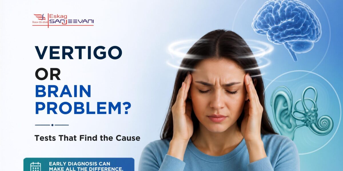
Vertigo is one of the most unsettling symptoms a person can experience. The room spins, balance disappears, and the fear of something serious sets in. But not every episode of spinning means there is a brain problem. In many cases, the cause is a minor inner ear issue that resolves with simple treatment. In others, vertigo is the first warning sign of a stroke, multiple sclerosis, or a brain tumour. Among adults over 65, the prevalence of dizziness reaches 30%, and in those over 75, it climbs to 50%. The key difference lies in getting the right vertigo test at the right time.
This blog explains how doctors distinguish an inner ear problem from a brain cause, which neurological tests are used, and when you need a specialist.
Key Takeaways
- Doctors may start with simple bedside tests such as the HINTS exam, Dix-Hallpike test and Romberg test to look for signs that may point to a central cause of vertigo.
- If a central cause is suspected, a brain MRI may be recommended. It can help doctors identify problems such as stroke or other changes in the brain.
- Peripheral causes like BPPV have an excellent outlook and respond well to repositioning manoeuvres; central causes require urgent investigation and targeted treatment of the underlying condition.
Quick Answer: A vertigo test identifies whether spinning comes from the inner ear or the brain using bedside exams and imaging.
Quick Links
- What Is Vertigo and Why Does It Happen?
- Peripheral vs Central Vertigo: Key Differences
- The Bedside Vertigo Test: How Doctors Start
- List of Neurological Tests for Vertigo Diagnosis
- Brain Problem Symptoms That Must Not Be Ignored
- How to Cure Vertigo Permanently: What the Evidence Says
- Who Is the Best Doctor for Vertigo?
What Is Vertigo and Why Does It Happen?
Vertigo is not simply feeling dizzy or light-headed. It is the sensation that you or your surroundings are spinning or rotating, even when you are completely still. It is a symptom of many underlying conditions rather than a disease in itself.
Vertigo arises when the vestibular system malfunctions. This system includes the inner ear, the vestibular nerve, the brainstem, and the cerebellum. When signals through this system are disrupted, the brain receives conflicting information about movement and position, producing the spinning sensation.
There are two broad categories:
Peripheral vertigo originates in the inner ear or vestibular nerve. It is the more common type and tends to carry a better outlook. Causes include benign paroxysmal positional vertigo (BPPV), Ménière’s disease, vestibular neuritis, and labyrinthitis [1].
Central vertigo originates in the brain or brainstem. It is less common but more serious. Causes include stroke, multiple sclerosis, brain tumours, and vestibular migraine. People with central vertigo often have severe instability and may be unable to walk without support.
The aetiology of dizziness is frequently multifactorial, involving degenerative vestibular changes, neurological disorders, and circulatory or musculoskeletal conditions. The most frequently diagnosed causes are BPPV, Meniere’s disease, vestibular neuritis, and central nervous system vascular diseases [2].
Also read: Comprehensive Neurology Services at Eskag Sanjeevani: Your Path to Better Brain Health.
Peripheral vs Central Vertigo: Key Differences
Before recommending any vertigo test, a doctor looks for distinguishing features between the two types. These differences guide every decision that follows.
| Feature | Peripheral Vertigo | Central Vertigo |
| Onset | Sudden, often positional | Gradual or sudden |
| Nystagmus direction | Unidirectional, horizontal-torsional | May reverse direction with gaze |
| Hearing loss or tinnitus | Often present | Usually absent |
| Neurological symptoms | Absent | Diplopia, dysarthria, ataxia may occur |
| Severity of imbalance | Mild to moderate | Often severe; cannot walk unaided |
| Nystagmus with gaze | Does not reverse direction | May reverse direction |
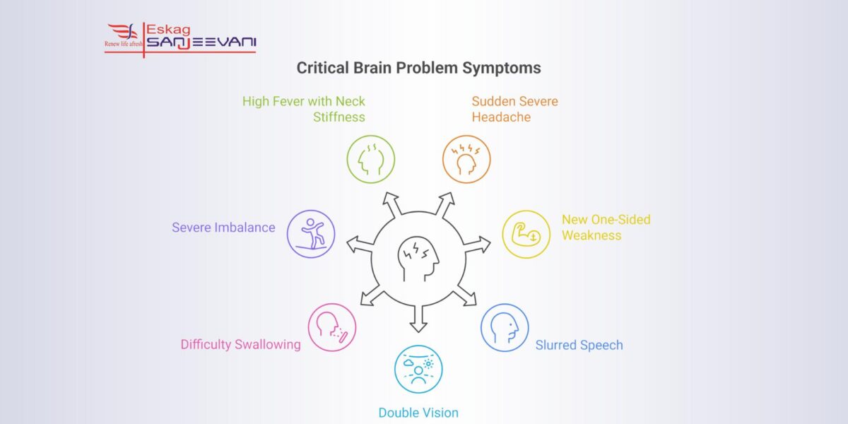
The presence of focal neurological symptoms alongside a vertigo problem, such as sudden severe headache, one-sided weakness, slurred speech, or double vision, is a red flag for a central cause. These brain problem symptoms warrant urgent evaluation and must not be dismissed.
The Bedside Vertigo Test: How Doctors Start
Most vertigo diagnoses begin with a focused clinical examination. The aim is to place the vertigo problem clearly in the peripheral or central category before any imaging is requested.
The HINTS Exam
The HINTS test (Head Impulse, Nystagmus, Test of Skew) is a three-part bedside assessment used in patients with acute, continuous vertigo. Research shows it may be more sensitive for detecting acute stroke than an MRI performed within the first 48 hours of symptom onset.
The three components work together:
- Head Impulse Test: The patient fixes their gaze on a target while the doctor rapidly rotates the head to one side. A corrective eye flick back to the target suggests a peripheral lesion. A normal response during active vertigo raises concern for a central cause.
- Nystagmus Assessment: In peripheral vertigo, the direction of nystagmus is consistent regardless of gaze direction. In central lesions, nystagmus may reverse direction when the patient looks in different directions.
- Test of Skew: The doctor covers one eye and then the other. If the uncovered eye moves up or down, it may suggest a problem in the brain.
- Looking at the results together: A normal head impulse test, direction-changing nystagmus and skew deviation can point to a central cause of vertigo and may require urgent assessment.
- Limitation to note: The HINTS exam is only reliable in patients with spontaneous, persistent nystagmus at the time of assessment.
The Dix-Hallpike Manoeuvre
This is the diagnostic test of choice when BPPV is suspected. The doctor turns the seated patient’s head 45 degrees to one side and then lowers them quickly to a lying position with the head slightly extended. A positive result produces vertigo with rotational nystagmus, confirming BPPV of the posterior semicircular canal.
Romberg’s Test
The patient stands with feet together, arms at their sides, and eyes closed. Significant unsteadiness or falling suggests dysfunction in the central nervous system or proprioceptive pathways [3].
Fukuda-Unterberger Test
The patient marches in place with eyes closed for 30 seconds [3]. Rotation or consistent drift to one side suggests an inner ear labyrinth problem on that side.
List of Neurological Tests for Vertigo Diagnosis
When the bedside examination raises clinical suspicion of a central cause, or when the vertigo problem is complex or recurrent, a vertigo specialist doctor will order formal investigations.
Brain MRI and MR Angiography
MRI is commonly used when doctors need to check for a central cause of vertigo. It visualises the posterior fossa, brainstem, and cerebellum with a level of detail that CT cannot match. CT scans have limited resolution in this region and should only be used when MRI is contraindicated or unavailable [1].
A case-control study of 232 vertigo patients and 232 controls found that white matter lesions were present in 39.2% of vertigo patients versus 25% of controls (p = 0.001). Lacunar lesions, Circle of Willis variations, and sinusitis were also significantly more frequent in the vertigo group. Following MRI, the most common diagnosis was BPPV (34.5%), followed by vertebrobasilar syndrome (11.6%) [2].
The standard brain MRI protocol for vertigo includes T1-weighted, T2-weighted, FLAIR, diffusion-weighted imaging (DWI), and a 3D FIESTA sequence specifically for inner ear evaluation. DWI is particularly critical for detecting acute cerebellar or brainstem infarcts, which can present as isolated vertigo with no other neurological signs.
Eskag Sanjeevani is equipped with MRI and 128-slice CT scanning for full neurological investigation, ensuring accurate diagnosis without delay.
Vestibular Test Battery
These are a set of objective tests used to assess vestibular function in the inner ear. They help determine whether vertigo is related to the inner ear or the brain. The tests may include:
- Videonystagmography (VNG): Uses video goggles to record eye movements during visual stimulation and changes in position. It can identify abnormal nystagmus.
- Caloric Testing: Warm or cool water or air is introduced into the ear canal to stimulate the inner ear. The resulting eye movements show how well each ear responds and whether the responses are balanced.
- Video Head Impulse Test (vHIT): Measures the vestibulo-ocular reflex using a high-speed camera, distinguishing peripheral from central disease in acute vertigo presentations.
- Vestibular Evoked Myogenic Potentials (VEMP): Measures neck and eye muscle responses to sound, assessing the utricle and saccule of the inner ear.
- Rotational Chair Testing: The patient sits in a motorised rotating chair wearing eye-tracking goggles, providing additional data on whether vertigo is of peripheral or central origin.
Audiometry and Tympanometry
A hearing check is a standard part of the vertigo workup. Unilateral hearing loss strongly points to a peripheral cause. Formal audiometry and tympanometry are more sensitive than bedside hearing tests for detecting sensorineural hearing loss and middle ear fluid. When a peripheral cause cannot be confirmed despite unilateral hearing loss, MRI with gadolinium contrast is recommended.
Auditory Brainstem Response (ABR)
Also called brainstem auditory evoked potentials, this test measures electrical signals along the auditory nerve to the brainstem. It objectively assesses the auditory pathway and is helpful when a tumour at the cerebellopontine angle, such as a vestibular schwannoma, is suspected.
Pre-MRI Vascular and Musculoskeletal Investigations
A case-control study found that carotid and vertebral artery ultrasound was the most common investigation performed before MRI, with atherosclerosis identified as the leading finding [2]. Cervical spine X-ray was also frequently used, with spondylosis found in the majority of positive cases. These pre-MRI assessments are essential for ruling out vascular and musculoskeletal contributors to a vertigo problem before proceeding to brain imaging.
Eskag Sanjeevani’s neurology department offers EEG, EMG, and NCV testing for cases where seizure-related dizziness or peripheral neuropathy is part of the differential diagnosis.
Brain Problem Symptoms That Must Not Be Ignored
Certain features alongside vertigo should prompt immediate emergency assessment. These brain problem symptoms suggest a central cause and cannot wait for a routine appointment:
- Sudden severe headache with no prior history, often described as the worst of one’s life
- New one-sided weakness or numbness in the face, arm, or leg
- Slurred or suddenly abnormal speech
- Double vision or abrupt loss of vision
- Difficulty swallowing or coordinating movement
- Severe imbalance with inability to stand or walk without falling
- High fever with neck stiffness, which may suggest meningitis
If any of these appear alongside vertigo, go to the emergency department without delay. Cerebellar stroke can closely mimic inner ear vertigo in its early hours, but its consequences are catastrophic if treatment is delayed. The stroke team at Eskag Sanjeevani is equipped and trained to respond immediately to these presentations.
How to Cure Vertigo Permanently: What the Evidence Says
Patients frequently ask how to cure vertigo permanently. The answer depends entirely on the underlying cause.
For BPPV, canalith repositioning manoeuvres such as the Epley manoeuvre dislodge displaced calcium crystals from the semicircular canals. This approach is the most commonly recommended intervention and can be performed in a clinic or taught for home use [1]. The case-control study confirmed physical training, including the Epley manoeuvre, as the most frequently applied treatment after MRI diagnosis.
For Ménière’s disease, a low-salt diet, diuretics, and vestibular rehabilitation can reduce episode frequency. Hearing loss from this condition may be permanent if it goes untreated for too long.
For vestibular neuritis, a short course of corticosteroids combined with early vestibular rehabilitation produces the best outcomes. The brain compensates through neuroplasticity over days to weeks.
For central causes such as stroke or multiple sclerosis, permanent resolution depends on treating the underlying condition. The most common pharmacological treatments used post-MRI include cognitive enhancers, anticoagulants, betahistine, and anxiolytics. Lifestyle counselling was the single most frequently applied intervention overall.
Prolonged use of vestibular suppressants such as antihistamines or benzodiazepines should be avoided. These medications interfere with the brain’s natural compensation mechanism and delay recovery.
Who Is the Best Doctor for Vertigo?
The right specialist depends on the suspected cause. A vertigo specialist doctor may be a neurologist when a central cause such as stroke, MS, or vestibular migraine is suspected, an ENT specialist when the vertigo appears to originate from the inner ear, or an audiologist for formal vestibular and hearing assessments.
In complex cases, specialists from neurology and ENT, radiology and physiotherapy may work together to assess and manage vertigo. A multidisciplinary approach can help when symptoms have more than one possible cause. Eskag Sanjeevani has neurology and ENT specialists who work across its Kolkata centres, with access to investigations such as MRI, CT, EEG, EMG and vestibular tests.
If you are unsure where to start, a general physician can perform the initial assessment and direct you to the best doctor for vertigo based on your specific symptom pattern.
Final Thoughts
Vertigo ranges from a benign positional inner ear problem to an early sign of serious brain disease. The right vertigo test, from the bedside HINTS exam to brain MRI and a full vestibular test battery, is the only reliable way to find the cause. Do not ignore sudden-onset vertigo that comes with neurological symptoms. Early, accurate diagnosis protects function and opens the path to the correct treatment. Book a consultation with Eskag Sanjeevani‘s neurology or ENT team if you are experiencing recurring or unexplained episodes of vertigo.

Microbiologist with over 2 years of experience in medical writing, specialising in evidence-based healthcare content.

Clinical experience in diagnosis, treatment, and evidence-based patient care across a range of conditions.
References
- Dy JS, Freeman AM. Vertigo in Clinical Practice: Evidence-Based Diagnosis and Treatment. In: StatPearls [Internet]. Treasure Island (FL): StatPearls Publishing; 2026 Jan-. Updated 2025 Sep 12. PMID: 29493978.
- Széphelyi K, Kóra S, Orsi G, Tollár J. Structural Brain Abnormalities, Diagnostic Approaches, and Treatment Strategies in Vertigo: A Case-Control Study. PMCID: PMC12472988. PMID: 41002934.
- Cleveland Clinic. Vertigo: Symptoms, Causes & Treatment. Reviewed 2023 May 9.
The HINTS bedside exam is highly accurate for ruling in or out a central cause in patients with acute continuous vertigo. For inner ear assessment, the vestibular test battery including VNG and caloric testing provides the most objective data. Brain MRI with DWI sequences is the gold standard when a stroke or structural brain lesion is suspected.
Yes, in some cases. Central vertigo caused by stroke, multiple sclerosis, or a brain tumour can present as spinning with little else initially. Brain problem symptoms such as sudden severe headache, one-sided weakness, slurred speech, or severe imbalance must be treated as a medical emergency and assessed immediately.
Brief positional episodes triggered by head movement are the classic pattern of BPPV, a benign inner ear condition. A neurologist or ENT specialist can often confirm this with the Dix-Hallpike test alone. A brain scan is not routinely needed for classic BPPV, but MRI is recommended if the Dix-Hallpike test is negative or neurological symptoms are present.
Duration varies by cause. BPPV episodes last seconds to a minute. Ménière’s attacks last 20 minutes to several hours. Vestibular neuritis produces continuous dizziness lasting days. BPPV responds very well to repositioning manoeuvres. There is no single answer to how to cure vertigo permanently, as treatment depends on identifying and addressing the specific underlying cause.
Start with a general physician or go directly to the neurology or ENT department of a multispecialty hospital. At Eskag Sanjeevani, vertigo patients are assessed by experienced neurologists and ENT specialists with access to on-site MRI, CT, EEG, EMG, and vestibular testing. Early specialist review prevents misdiagnosis and reduces the risk of complications from an undetected central cause.